

Cavoatrial junction
Images
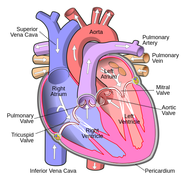
Cavoatrial junction
Defining the Cavoatrial Junction
The cavoatrial junction (CAJ) represents a fundamental anatomical boundary within the thoracic vasculature, signifying the confluence of the superior vena cava (SVC) with the superior aspect of the right atrium (RA). This junction is not merely a point of contact but a transition zone where the venous wall integrates into the atrial myocardium. The SVC, a large deoxygenated blood conduit originating from the brachiocephalic veins, terminates at this junction, marking the inferior extent of the SVC as a distinct anatomical entity.
Below this point, the vessel is considered part of the RA. Understanding this precise demarcation is essential for accurate anatomical description and for interpreting diagnostic imaging. The CAJ's significance extends beyond basic anatomy; it is a critical reference point for hemodynamic monitoring and the safe administration of therapies that require central venous access, influencing how blood flow dynamics are understood and managed within the cardiopulmonary system.
Its definition underscores the intricate structural continuity between the venous system and the heart's primary receiving chamber.
Radiographic Localization
Accurate localization of the cavoatrial junction is paramount for safe medical interventions, particularly central venous catheterization. Radiographic identification relies on specific anatomical landmarks rather than subjective visual cues. The most reliable method involves identifying the carina, the bifurcation of the trachea, and then referencing the overlying thoracic vertebrae.
The CAJ is typically found two vertebral body units inferior to the carina, crucially including the intervertebral disc space between these two vertebrae but excluding the disc space below the second vertebra. This methodical counting ensures a consistent and reproducible landmark identification. Traditional methods, such as identifying a 'bulge' in the cardiac silhouette on a chest X-ray, are considered unreliable due to the potential for displacement caused by cardiac enlargement, pulmonary pathology, or patient positioning.
Advanced imaging techniques and electrocardiographic monitoring further refine the confirmation of catheter tip placement relative to this critical junction.
Clinical Significance
The cavoatrial junction holds profound clinical importance, primarily in the context of central venous catheterization. Patients requiring long-term or intensive therapies, such as chemotherapy, parenteral nutrition, or vasopressors, often necessitate central venous access. Central venous catheters, when inserted via the upper body, are ideally positioned with their tips at or just superior to the CAJ within the SVC.
This strategic placement ensures that infusates, particularly those that are hyperosmolar or potentially vesicant, are rapidly diluted in the large volume of blood entering the RA. This dilution minimizes the risk of local endothelial damage to the vessel wall and reduces the potential for phlebitis or thrombosis. The CAJ serves as a critical anatomical target that balances the need for effective drug delivery with the imperative to avoid direct irritation or injury to the heart's delicate structures, thereby optimizing patient safety and therapeutic efficacy.
Catheter Tip Placement
The precise positioning of central venous catheter tips relative to the cavoatrial junction remains a subject of nuanced clinical practice and ongoing discussion. While placement within the SVC just above the CAJ is widely accepted for minimizing local irritation, some practitioners advocate for positioning the tip slightly within the RA, proximal to the CAJ. The rationale behind avoiding the CAJ itself stems from concerns about potential cardiac arrhythmias or even cardiac tamponade if the catheter tip encroaches too deeply into the RA or right ventricle.
These complications arise from mechanical irritation of the heart's conduction system or epicardium. Conversely, others argue that modern catheter materials and verification methods, such as electrocardiogram (ECG) monitoring which detects characteristic changes in electrical activity correlating with tip proximity to the heart, demonstrate that RA placement is safe and may offer advantages in certain clinical scenarios. Chest X-rays continue to be a valuable tool for confirming tip position, especially when ECG monitoring is contraindicated or inconclusive, underscoring the multi-modal approach to ensuring safe and effective central venous access.
See also
Based on content from Wikipedia · Licensed under CC BY-SA 4.0