

Bleeding: When Your Body Leaks!
Images
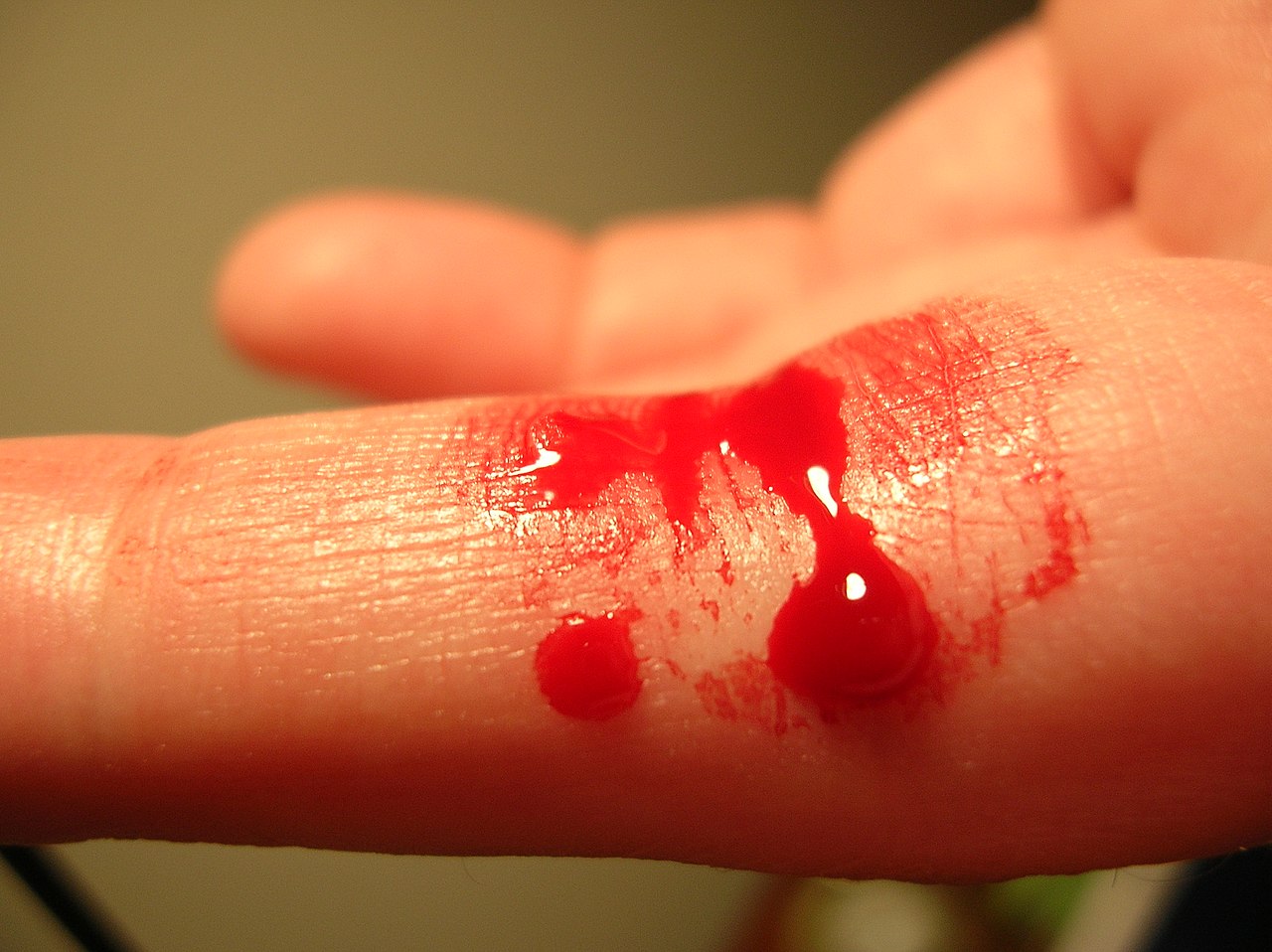
Bleeding
The Complex Cascade of Hemostasis
Hemostasis is the body's sophisticated process for stopping bleeding. It's a tightly regulated cascade involving multiple components, initiated immediately upon vascular injury. The primary goal is to form a stable fibrin clot at the site of damage, preventing excessive blood loss while maintaining blood flow elsewhere.
This process begins with primary hemostasis: vasoconstriction, where blood vessels narrow to reduce blood flow, followed by platelet aggregation. Platelets adhere to exposed collagen in the damaged vessel wall, facilitated by von Willebrand factor (vWF). They then activate, change shape, and aggregate, forming a temporary platelet plug.
Secondary hemostasis involves the coagulation cascade, a series of enzymatic reactions involving plasma proteins called clotting factors. This cascade culminates in the conversion of soluble fibrinogen into insoluble fibrin, which reinforces the platelet plug, creating a robust and stable clot. This intricate interplay ensures rapid and effective sealing of injured vessels.
Mechanisms of Vascular Injury and Pathological Bleeding
Vascular injury can occur through various mechanisms, leading to bleeding. Traumatic injuries, such as cuts, abrasions, and blunt force trauma, directly breach the integrity of blood vessels. Surgical procedures inherently involve controlled vascular transection, necessitating meticulous hemostatic techniques.
Pathological conditions can also compromise vascular integrity. For instance, inflammatory processes can weaken vessel walls, making them more prone to rupture. Certain diseases, like aneurysms, involve localized dilations of blood vessels that are susceptible to rupture.
Furthermore, bleeding disorders, such as hemophilia or von Willebrand disease, represent inherited defects in the hemostatic pathway, leading to spontaneous or exaggerated bleeding even with minor injuries. Understanding the specific mechanism of injury is crucial for effective management and prevention of excessive blood loss.
Evolution of Hemostatic Management
The management of bleeding has evolved dramatically throughout history. Ancient civilizations employed rudimentary methods like direct pressure, ligation of vessels, and cauterization. The development of surgical techniques in the Renaissance and Enlightenment periods saw increased focus on controlling bleeding during procedures, though often with high mortality rates due to infection and uncontrolled hemorrhage.
The 19th century marked a turning point with the introduction of anesthesia, antisepsis, and the understanding of germ theory, significantly improving surgical outcomes. The discovery of blood groups and the development of blood transfusion techniques in the early 20th century provided a revolutionary way to replace lost blood volume. Today, hemostatic management encompasses a wide array of sophisticated tools and therapies, including advanced surgical sealants, topical hemostatic agents, recombinant clotting factors, and minimally invasive interventional radiology techniques, offering precise and effective control over bleeding in diverse clinical scenarios.
Therapeutic Interventions and Future Directions in Bleeding Control
Modern medicine offers a comprehensive armamentarium for managing bleeding. For minor injuries, direct pressure and topical agents are often sufficient. In surgical settings, electrocautery, ultrasonic scalpels, and various hemostatic dressings (e.g., gelatin sponges, oxidized cellulose) are employed.
For more severe or systemic bleeding, blood transfusions remain a cornerstone, providing essential clotting factors and red blood cells. Pharmacological agents, such as desmopressin (DDAVP) for certain platelet disorders or tranexamic acid to inhibit fibrinolysis, are also critical. For patients with inherited bleeding disorders, replacement therapy with specific clotting factors is standard. Future directions in bleeding control research include the development of novel biomaterials that promote rapid and robust clot formation, targeted drug delivery systems to activate hemostatic pathways precisely where needed, and advanced imaging techniques for real-time monitoring of hemostasis during complex procedures.
Go deeper
Frequently Asked Questions
Why does a scraped knee turn red and bleed?+
How does your body stop bleeding after an injury?+
What are platelets and what do they do when you get a cut?+
Why do some people bleed more easily, like those with hemophilia?+
How do doctors help stop bleeding during surgery?+
Based on content from Wikipedia · Licensed under CC BY-SA 4.0