

Vesicouterine Pouch
Images
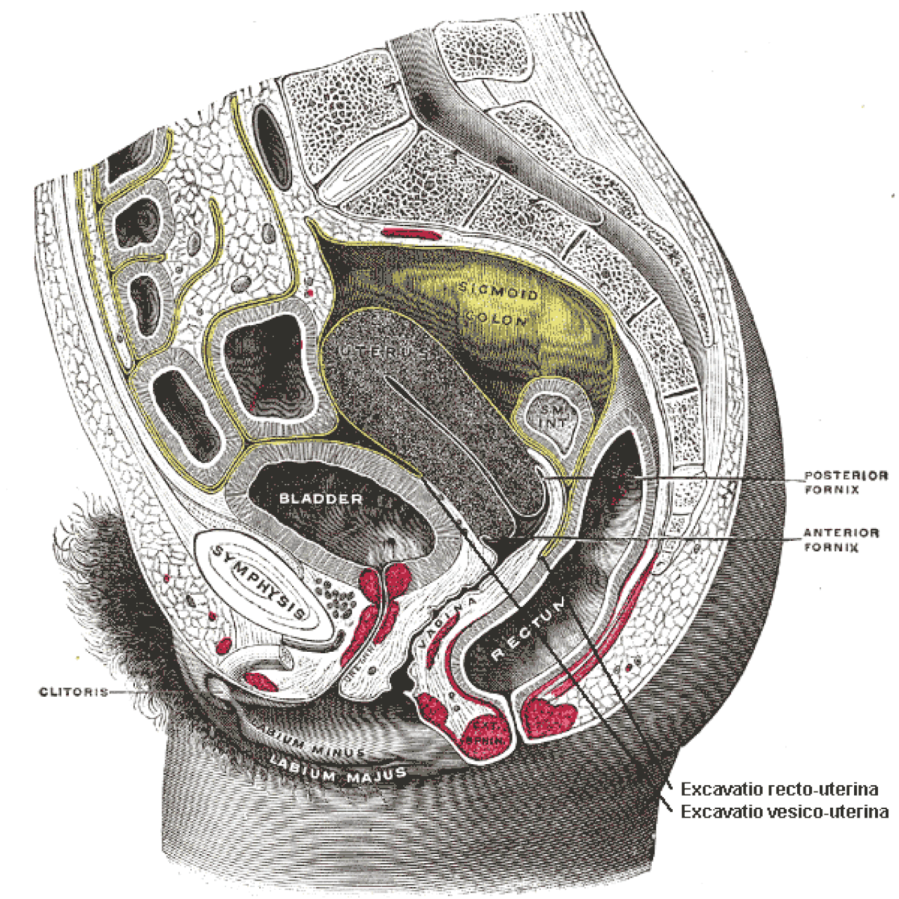
Vesicouterine pouch
Formation and Anatomical Context of the Vesicouterine Pouch
The vesicouterine pouch, also known as the uterovesicle pouch, represents a specific anatomical landmark within the female pelvic cavity. It is not an intrinsic organ but rather a dependent space formed by the reflection of the peritoneum. This serous membrane, the peritoneum, lines the abdominal cavity and invests the pelvic viscera.
In the female pelvis, the peritoneum drapes over the superior surface of the urinary bladder and then reflects posteriorly onto the anterior surface of the uterus, creating this shallow recess. Its anterior boundary is the posterior aspect of the bladder, and its posterior boundary is the anterior aspect of the uterus. It is consistently shallower and situated more anteriorly than the rectouterine pouch (pouch of Douglas), which lies between the uterus and the rectum.
This specific configuration is a consequence of the developmental anatomy of the pelvic organs and the way the peritoneum envelops them. The relative shallowness is a key distinguishing feature, influencing its clinical behavior.
Functional Implications and Anatomical Relationships
While the vesicouterine pouch is a passive space, its existence has functional implications related to the spatial organization and support of pelvic organs. It helps to maintain a distinct separation between the urinary bladder and the uterus, preventing undue friction or pressure between these two organs during their physiological functions, such as bladder filling and uterine distension. This anatomical relationship is crucial for the normal functioning of both the urinary and reproductive systems.
The pouch's position also influences the distribution of intra-abdominal pressure and the mobility of these organs. Understanding its precise location is fundamental for appreciating the intricate biomechanics of the female pelvis and how organs interact within this confined space. It contributes to the overall compartmentalization and support provided by the peritoneal lining.
Clinical Significance
The vesicouterine pouch holds considerable clinical significance, particularly in diagnostic imaging and surgical interventions. As a dependent space within the peritoneal cavity, it is prone to accumulating free fluid, whether serous fluid, blood, or pus, in various pathological conditions. Conditions such as ascites (fluid in the abdomen), pelvic inflammatory disease, post-operative complications, or intraperitoneal hemorrhage can lead to fluid collection in this pouch, making it a target for detection via ultrasound, CT, or MRI scans.
Radiologists and clinicians often assess the presence and characteristics of fluid within the vesicouterine pouch as part of a comprehensive pelvic examination. Furthermore, for surgeons performing gynecological, urological, or general abdominal procedures in the pelvic region, precise knowledge of the vesicouterine pouch's boundaries is essential for safe dissection and to avoid inadvertent injury to the bladder, uterus, or surrounding vasculature and nerves. It serves as a critical anatomical landmark during laparoscopic and open surgeries.
Comparative Anatomy and Pathological Variations
Comparing the vesicouterine pouch to the rectouterine pouch highlights differences in their depth and clinical implications. The deeper rectouterine pouch is more likely to collect significant amounts of fluid and is often the primary site for fluid accumulation in conditions like ascites. The vesicouterine pouch, being shallower, may collect less fluid but is still a relevant space.
Pathological variations can include adhesions forming within the pouch due to inflammation or previous surgery, potentially altering organ mobility and contributing to conditions like infertility or chronic pelvic pain. In rare congenital anomalies or during significant organ displacement, the typical configuration of the vesicouterine pouch might be altered. Its consistent presence and predictable location, however, make it a reliable reference point in both normal and pathological anatomy.
See also
Frequently Asked Questions
What is the vesicouterine pouch?+
Where is the vesicouterine pouch located?+
Why is the vesicouterine pouch important for doctors and surgeons?+
How can doctors find fluid in the vesicouterine pouch?+
How is the vesicouterine pouch different from the rectouterine pouch?+
Based on content from Wikipedia · Licensed under CC BY-SA 4.0