

The Pineal Gland: Your Brain's Tiny Sleep Helper!
Images

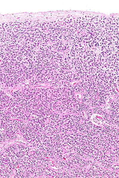
Capillary Supply in Young Human Pineal Gland




Anatomy and Histology
The pineal gland, also known as the epiphysis cerebri, is a small, pinecone-shaped endocrine organ situated in the epithalamus of the vertebrate brain. It is strategically located near the center of the brain, nestled within a groove where the two thalami converge, posterior to the habenular commissure. This location places it within the epithalamus, a region involved in regulating sleep-wake cycles and emotional responses.
Histologically, the pineal gland is composed primarily of pinealocytes, which are responsible for synthesizing melatonin, and glial cells, predominantly astrocytes. It is classified as a circumventricular organ, characterized by fenestrated capillaries that allow for relatively free exchange of molecules between the gland's tissue and the bloodstream, facilitating efficient hormone distribution.
Evolutionary Trajectory
The evolutionary history of the pineal gland suggests a fascinating transformation. In many lower vertebrates and even some invertebrates, homologous structures function as direct light-sensing organs, often referred to as the parietal eye or pineal eye. These structures can detect light intensity and duration, influencing behaviors like thermoregulation and migration.
Reconstructions of evolutionary patterns indicate that the pineal gland in mammals and birds likely originated from an atrophied photoreceptor. Over eons, this primitive light-sensing organ evolved into a sophisticated neuroendocrine organ, losing its direct light-detecting capability but retaining its crucial role in translating environmental light cues into hormonal signals.
The Melatonin Synthesis Pathway and Circadian Control
The pineal gland's primary function is the synthesis and secretion of melatonin, a hormone derived from the amino acid tryptophan via serotonin. This process is tightly regulated by the suprachiasmatic nucleus (SCN) of the hypothalamus, the master circadian pacemaker. Light perceived by the retina is transmitted via the retinohypothalamic tract to the SCN, which then signals the pineal gland through a multi-synaptic pathway involving the sympathetic nervous system.
In darkness, norepinephrine is released, activating beta-adrenergic receptors on pinealocytes, which upregulates the expression and activity of key enzymes in the melatonin synthesis pathway, including arylalkylamine N-acetyltransferase (AA-NAT) and hydroxyindole-O-methyltransferase (HIOMT). Melatonin is released into the bloodstream, acting on its receptors throughout the body to promote sleep onset, reduce alertness, and synchronize peripheral circadian clocks.
Historical Perspectives and Philosophical Interpretations
The pineal gland has been a subject of scientific and philosophical inquiry for millennia. Ancient Greek physicians like Galen recognized its presence but were uncertain of its function, likening its shape to a pinecone and suggesting it might serve as a structural support. The name 'pineal' itself derives from the Latin 'pinealis,' meaning pinecone-shaped. During the Renaissance, its enigmatic nature led to more speculative theories.
Most notably, René Descartes, in the 17th century, proposed the pineal gland as the 'principal seat of the soul' and the primary site of mind-body interaction, a philosophical concept that highlighted its perceived centrality and uniqueness within the brain. While modern neuroscience has elucidated its physiological role, Descartes' ideas underscore the historical intrigue surrounding this small but significant organ.
Clinical Significance and Modern Relevance
Disruptions in pineal gland function and melatonin production are linked to various health issues. Seasonal Affective Disorder (SAD), a type of depression related to changes in seasons, is thought to be influenced by altered melatonin cycles due to reduced light exposure. Sleep disorders, such as insomnia, can also be associated with dysregulated melatonin rhythms.
Furthermore, the pineal gland's role in regulating circadian rhythms extends to broader physiological processes, including immune function, metabolism, and even cancer progression, as melatonin exhibits oncostatic properties. Research into exogenous melatonin supplementation and light therapy continues to explore therapeutic interventions for these conditions, underscoring the pineal gland's ongoing importance in human health and well-being.
See also
Frequently Asked Questions
What is the pineal gland and where is it in my brain?+
How does the pineal gland help me sleep?+
Why does the pineal gland need light to work?+
What cells make melatonin in the pineal gland?+
Did the pineal gland always look like a pinecone?+
Based on content from Wikipedia · Licensed under CC BY-SA 4.0