

Microscope: The Amazing Magnifying Glass!
Images

Microscope


The Genesis of Microscopic Vision
The invention of the microscope was not a singular eureka moment but rather an evolution rooted in the development of optics. While the exact origins are debated, the most widely accepted account credits Dutch spectacle makers Zacharias and Hans Janssen with creating one of the first compound microscopes around 1590. Their innovation involved combining multiple lenses within a tube, a principle that would define microscopy for centuries.
Early microscopes were rudimentary, offering limited magnification and often producing distorted images. However, they represented a profound leap in human perception, allowing for the first glimpses into a previously invisible world. Figures like Antonie van Leeuwenhoek, in the 17th century, significantly advanced the field by developing powerful single-lens microscopes, meticulously grinding his own lenses to achieve remarkable clarity and magnification, leading him to discover microorganisms, sperm cells, and blood flow in capillaries.
These early pioneers laid the groundwork for the sophisticated instruments we use today.
The Indispensable Role of Microscopy in Scientific Advancement
Microscopes are foundational tools across virtually every scientific discipline, acting as gateways to understanding complexity at its most fundamental level. In biology and medicine, they are critical for cell biology, enabling the study of cellular structures, processes like mitosis and meiosis, and the identification of pathogens responsible for diseases. This has led to breakthroughs in diagnostics, therapeutics, and our understanding of life itself.
Materials science relies heavily on microscopy to analyze the microstructures of metals, polymers, and ceramics, informing the development of new materials with enhanced properties for engineering and manufacturing. In fields such as forensic science, microscopy is used to examine trace evidence, like fibers or hair, providing crucial links in investigations. The ability to visualize and analyze these minute details is not merely observational; it drives hypothesis formation, experimental design, and ultimately, scientific discovery and innovation.
The Mechanics of Magnification
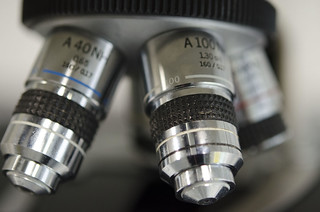
Microscopes achieve magnification through the manipulation of light or electrons. Light microscopes, the most common type, utilize lenses to refract (bend) light that passes through or reflects off a specimen. A compound light microscope typically employs an objective lens and an eyepiece lens, each magnifying the image sequentially.
The resolution, or the ability to distinguish between two closely spaced points, is limited by the wavelength of visible light. Electron microscopes, developed later, overcome this limitation by using beams of electrons, which have much shorter wavelengths. Transmission Electron Microscopes (TEMs) pass electrons through an ultra-thin specimen, revealing internal structures with extremely high resolution, while Scanning Electron Microscopes (SEMs) scan the surface of a specimen with electrons, producing detailed 3D images.
Each type of microscopy offers unique advantages for visualizing different aspects of the microscopic world.
Beyond the Visible
The evolution of microscopy continues with increasingly sophisticated techniques that push the boundaries of what can be observed. Super-resolution microscopy, for example, employs clever optical tricks to bypass the diffraction limit of light, achieving resolutions far beyond traditional light microscopes. This allows scientists to visualize molecular processes within living cells in real-time.
Confocal microscopy uses pinholes to eliminate out-of-focus light, creating sharp, optical sections of specimens, which can then be reconstructed into 3D images. Furthermore, the integration of microscopy with other technologies, such as spectroscopy and artificial intelligence, is opening new avenues for analysis and discovery. Future frontiers may involve even more advanced imaging techniques, potentially allowing us to visualize atomic-level structures in biological systems or explore the quantum realm of matter.
Microscopes in Action
The practical applications of microscopes permeate modern society. In healthcare, they are indispensable for pathology labs, where tissue samples are examined for signs of disease, and for hematology, where blood cells are analyzed. In the pharmaceutical industry, microscopes are used to study drug interactions with cells and to ensure the quality of manufactured medicines.
In manufacturing, they are vital for quality control, inspecting the integrity of microelectronic components, the surface finish of materials, and the structure of alloys. Even in fields like archaeology and art conservation, microscopes help in analyzing ancient artifacts and artworks to understand their composition and condition. The microscope is a silent but powerful partner in countless industries, ensuring precision, safety, and innovation.
Go deeper
Frequently Asked Questions
What is a microscope?+
Who invented the first microscope?+
How do microscopes make things look bigger?+
Why do scientists use microscopes?+
What is the difference between a light microscope and an electron microscope?+
Based on content from Wikipedia · Licensed under CC BY-SA 4.0