

Lithopedion: The Amazing Stone Baby!
Images
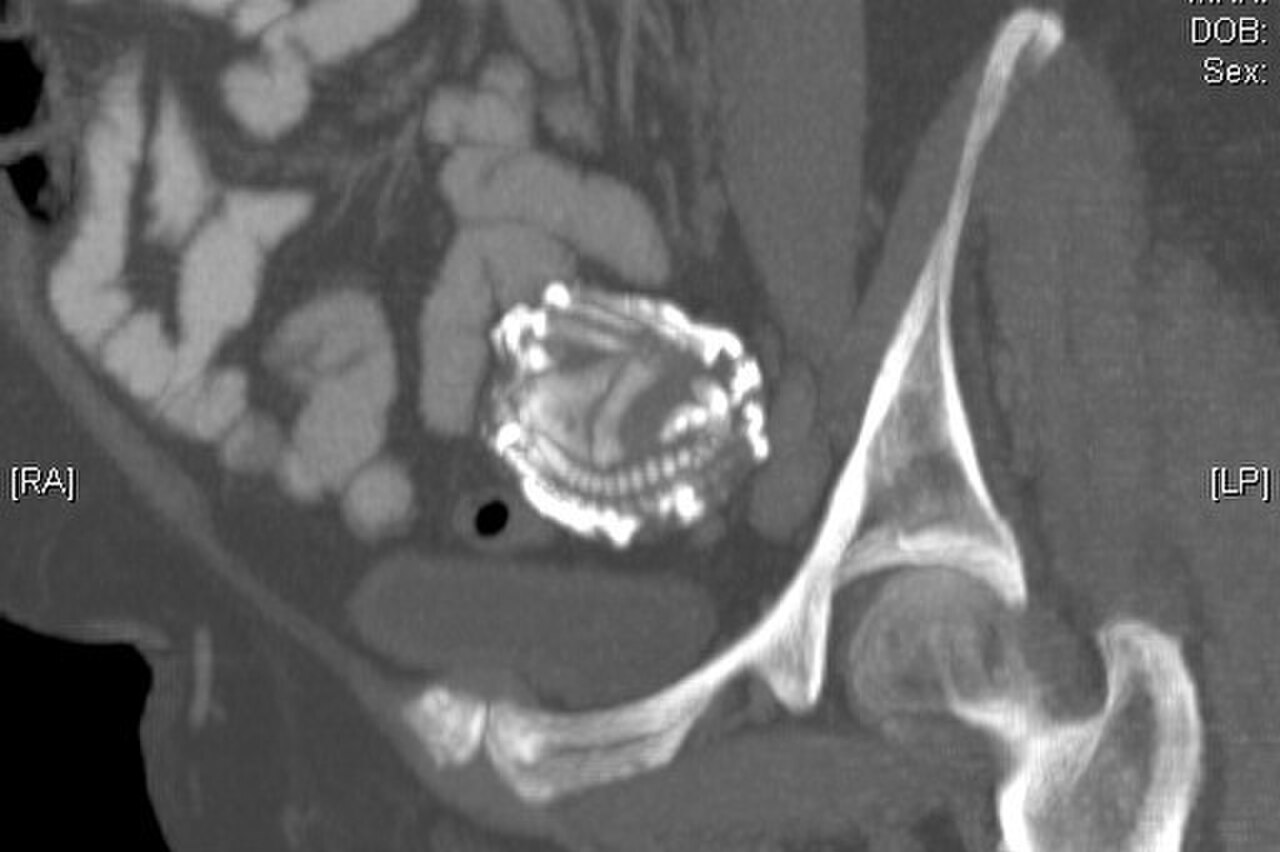
File:Incidental lithopedian upon TAH 50 year.jpg


The Genesis of a Stone Infant
A lithopedion, a term derived from the Greek 'lithos' (stone) and 'paidion' (infant), represents an exceptionally rare and complex medical outcome. It arises from an abdominal pregnancy, a life-threatening form of ectopic pregnancy where the fertilized ovum implants and develops within the peritoneal cavity rather than the uterine cavity. This occurs in approximately 1 in 11,000 pregnancies, but only a minuscule fraction, between 1.5% and 1.8%, progress to lithopedion formation.
The critical factor is the death of the fetus, typically due to insufficient blood supply or mechanical complications. If the fetus is too large to be resorbed by the maternal tissues and cannot be expelled, the body initiates a foreign body reaction. This involves the slow, progressive deposition of calcium salts, primarily from the mother's bloodstream, around the deceased fetus.
This calcification creates a hard, protective shell, effectively mummifying the fetus and isolating it from the maternal system. This process prevents sepsis and further inflammatory responses, acting as a biological containment strategy. The calcification can begin as early as 14 weeks gestation and continue for years, resulting in a fully encased fetal form.
A Decades-Long Secret
One of the most striking aspects of lithopedia is the prolonged period during which they can remain undiagnosed. It is not uncommon for women to carry a lithopedion for decades, often well past menopause. The mean age at diagnosis, according to a review of 128 cases by T.S.P.
Tien, was 55 years, with the oldest recorded case being a woman of 100 years. The average duration of carriage before diagnosis was 22 years, and remarkably, some women carried their lithopedion for over 50 years. Diagnosis is frequently incidental, occurring when a patient undergoes radiographic imaging, such as X-rays or CT scans, for unrelated medical conditions.
Symptoms, if present, are often vague and may include chronic abdominal pain, discomfort, or a palpable mass. The discovery can be a profound shock, both medically and psychologically, for the patient and clinicians alike, given the rarity and the long silent presence of the calcified fetus.
The Rarity and Significance
The sheer rarity of lithopedia makes them a subject of significant medical interest. With only approximately 300 documented cases in over 400 years of medical literature, each instance provides valuable insights into the body's adaptive and defensive capabilities under extreme circumstances. The significance extends beyond mere curiosity; it underscores the potential for unusual pregnancy outcomes and the body's remarkable capacity to manage and contain pathological entities.
The fact that women can carry a lithopedion for such extended periods and, in several documented cases, subsequently achieve successful pregnancies and deliver healthy children, highlights the resilience of the female reproductive system and the body's ability to compartmentalize and tolerate significant internal abnormalities. This phenomenon challenges our understanding of pregnancy complications and the long-term effects of ectopic gestations.
Beyond the Stone
The management of a lithopedion typically involves surgical removal, especially if it is causing symptoms or if there is concern about potential complications. However, if asymptomatic, conservative management might be considered, though surgical intervention is often preferred for definitive resolution. Related conditions include other forms of ectopic pregnancy, such as tubal pregnancies, and rare instances of fetal lithopedion in animals.
In modern medicine, advanced imaging techniques like MRI and CT scans have improved diagnostic accuracy, allowing for earlier detection and better surgical planning. The study of lithopedia also contributes to our broader understanding of calcification processes in the body, which has implications for conditions like atherosclerosis and kidney stones. While a lithopedion is a rare and often startling discovery, it serves as a powerful reminder of the intricate and sometimes astonishing ways the human body can respond to extraordinary challenges.
See also
Frequently Asked Questions
What is a lithopedion?+
How does a lithopedion form?+
Why can a lithopedion stay inside a woman for many years?+
Can a woman have babies after having a lithopedion?+
How do doctors discover a lithopedion?+
Based on content from Wikipedia · Licensed under CC BY-SA 4.0