

Ewing's sarcoma
Images

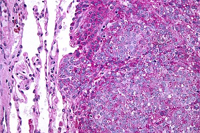
Ewing sarcoma of the chest wall




The Cellular Genesis of Ewing's Sarcoma
Ewing's sarcoma is a highly aggressive malignant neoplasm characterized by the uncontrolled proliferation of primitive neuroectodermal cells. It primarily affects bone and soft tissues, with a predilection for the appendicular skeleton, pelvis, and trunk. The hallmark of Ewing's sarcoma at the molecular level is the presence of specific chromosomal translocations, most commonly t(11;22)(q24;q12), which fuses the EWSI gene on chromosome 11 with the FLI1 gene on chromosome 22.
This fusion creates a chimeric transcription factor, EWS-FLI1, that dysregulates gene expression, promoting cellular growth, survival, and invasiveness. Other less common translocations, such as t(21;22) involving EWSI and ERG, also occur. These genetic alterations are considered driver mutations, initiating the oncogenic process.
The tumor cells themselves are small, round, blue cells with scant cytoplasm and indistinct borders, often exhibiting areas of necrosis and hemorrhage, reflecting their rapid growth and vascular compromise.
From Obscurity to Recognition
The entity now known as Ewing's sarcoma was first described in 1864 by the American physician James Ewing. However, his initial description was of a primary bone tumor in the long bones, and it wasn't until the mid-20th century that its distinct clinicopathological and radiological features were fully elucidated. Initially, it was often confused with other bone tumors like osteosarcoma or reticulum cell sarcoma.
The development of immunohistochemistry and electron microscopy in the latter half of the 20th century was pivotal in distinguishing Ewing's sarcoma from other small round blue cell tumors, particularly by identifying markers like CD99. The understanding of its neuroectodermal origin also evolved significantly. This historical progression highlights the scientific journey from initial observation to precise classification and molecular understanding, crucial for accurate diagnosis and treatment development.
The Critical Imperative
Ewing's sarcoma represents a significant challenge in pediatric and young adult oncology, accounting for approximately 10-15% of all primary bone cancers. Its aggressive nature and propensity for early metastasis, often to the lungs and bone marrow, contribute to a substantial morbidity and mortality rate. Therefore, a deep understanding of its biology is paramount for developing effective therapeutic strategies. Research into Ewing's sarcoma drives advancements in cancer biology, particularly in understanding gene regulation, cell signaling pathways, and the tumor microenvironment.
Furthermore, the development of multimodal treatment approaches, combining surgery, chemotherapy, and radiation, has improved survival rates, but significant unmet needs remain, especially for patients with metastatic or recurrent disease. Continued investigation is vital for improving long-term outcomes and reducing treatment-related toxicities.
Unraveling the Mechanism
The pathophysiology of Ewing's sarcoma is intrinsically linked to the aberrant expression of the EWS-FLI1 fusion protein. This chimeric transcription factor acts as a master regulator, hijacking normal cellular processes. It influences genes involved in cell proliferation, differentiation, apoptosis (programmed cell death), and angiogenesis (the formation of new blood vessels).
EWS-FLI1 can activate genes that promote cell cycle progression, such as Cyclin D1, and inhibit tumor suppressor genes. It also plays a role in promoting epithelial-mesenchymal transition (EMT), a process that enhances cell motility and invasiveness, facilitating metastasis. The tumor microenvironment, including immune cells and stromal components, also plays a complex role in tumor progression and response to therapy.
Understanding these intricate molecular mechanisms is key to identifying novel therapeutic targets.
Anatomical Distribution and Diagnostic Considerations
Ewing's sarcoma exhibits a characteristic anatomical distribution. The diaphysis (shaft) of long bones, particularly the femur, tibia, and humerus, are the most common sites. The pelvis is also a frequent location, followed by the ribs and spine.
Extraskeletal Ewing's sarcoma, arising in soft tissues, can occur anywhere but is most often found in the paravertebral region, chest wall, and abdomen. Diagnosis relies on a combination of clinical presentation, imaging studies (X-ray, MRI, CT, bone scan, PET scan), and histopathological examination of a biopsy specimen. Radiographically, it often appears as a destructive lesion with periosteal reaction (e.g., Codman's triangle or onion-skinning).
Definitive diagnosis requires immunohistochemical staining for markers like CD99, FLI1, and NKX2.2, alongside molecular testing for the characteristic translocations, which are crucial for confirming the diagnosis and guiding treatment.
See also
Frequently Asked Questions
What is Ewing's sarcoma?+
Why does Ewing's sarcoma grow so quickly?+
Where does Ewing's sarcoma usually appear in the body?+
How do doctors find out if a tumor is Ewing's sarcoma?+
What treatments do doctors use for Ewing's sarcoma?+
Based on content from Wikipedia · Licensed under CC BY-SA 4.0