

CT Scan
Images
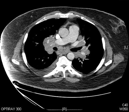
Sarcoidosis - Chest CT scan Case 242


The Genesis of CT
The development of computed tomography (CT) in the early 1970s marked a paradigm shift in medical imaging. Prior to CT, physicians primarily relied on two-dimensional projection radiography (standard X-rays) and more invasive procedures like angiography. The concept of reconstructing a 3D image from multiple 2D projections was theorized earlier, but it was the convergence of advancements in X-ray detection, computer processing power, and sophisticated mathematical algorithms that made CT a reality.
Allan Cormack, a physicist, and Godfrey Hounsfield, an electrical engineer, independently pioneered the mathematical principles and practical implementation, respectively. Their collaborative efforts, recognized by the 1979 Nobel Prize, laid the foundation for a technology that could visualize internal structures with unprecedented detail and clarity, moving beyond simple shadowgrams to true cross-sectional anatomy.
The Mechanics of CT
A modern CT scanner is a marvel of engineering. At its core is a gantry housing a rotating X-ray tube and an array of detectors. As the patient lies on a motorized table that moves through the gantry's opening (the 'donut'), the X-ray tube emits a fan-shaped beam of X-rays.
This beam rapidly rotates around the patient, passing through the body at numerous angles. Different tissues attenuate the X-rays to varying degrees based on their density and composition. The detectors on the opposite side capture the transmitted X-rays, measuring the intensity of the beam after it has passed through the body.
These raw measurements, known as projections, are then transmitted to a powerful computer. Using complex tomographic reconstruction algorithms, such as filtered back-projection or iterative reconstruction, the computer processes these thousands of data points to create detailed cross-sectional images, or 'slices,' of the body. These slices can then be digitally stacked and reformatted to create 3D reconstructions.
The Indispensable Role of CT in Modern Medicine
CT scans are a cornerstone of modern medical diagnosis and management due to their speed, availability, and diagnostic accuracy across a wide spectrum of conditions. They are invaluable in emergency medicine for rapidly assessing trauma patients, identifying internal bleeding, organ damage, or fractures. In neurology, CT is crucial for detecting strokes, brain hemorrhages, and tumors.
Oncologists use CT to detect, stage, and monitor various cancers, as well as to guide biopsies and radiation therapy. The ability to visualize bone structures makes CT essential for diagnosing complex fractures and planning orthopedic surgeries. Furthermore, CT scans are often preferred over MRI for patients with metallic implants or pacemakers, as the strong magnetic fields in MRI can be dangerous for these individuals.
This versatility ensures CT remains a vital tool in virtually every medical specialty.
Beyond Diagnosis
While CT's primary role is diagnostic, its applications extend further. CT angiography (CTA) uses contrast agents to visualize blood vessels, aiding in the detection of aneurysms, dissections, and blockages. CT-guided interventions, such as biopsies and drain placements, allow for precise targeting of lesions, minimizing risk and improving success rates.
The technology continues to evolve, with advancements like dual-energy CT providing more information about tissue composition, and photon-counting detectors promising higher resolution and reduced radiation dose. The integration of artificial intelligence (AI) is also transforming CT, with algorithms assisting in image analysis, anomaly detection, and workflow optimization. Despite the rise of other imaging modalities, CT's speed, broad applicability, and ongoing technological improvements ensure its continued relevance and importance in healthcare.
See also
Frequently Asked Questions
What is a CT scan and how does it work?+
Why do doctors use CT scans instead of regular X‑rays?+
How can a CT scan help in emergencies?+
Why is a CT scan better for people with metal implants?+
What is CT angiography and what does it show?+
Based on content from Wikipedia · Licensed under CC BY-SA 4.0