

Chromatophores: Nature's Tiny Color Changers!
Images
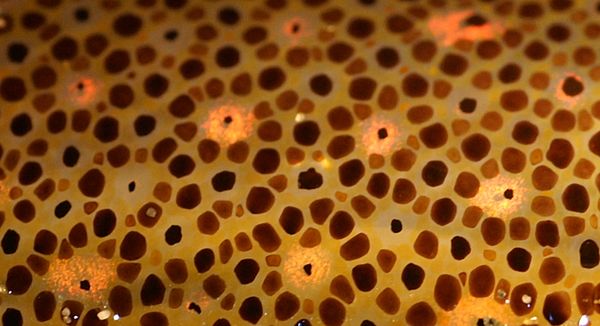
Chromatophore


The Cellular Architecture of Animal Coloration
Chromatophores represent a sophisticated cellular adaptation found across a wide spectrum of ectothermic animals, including fish, amphibians, reptiles, and cephalopods. These cells are the primary effectors of skin and eye coloration, functioning through two main principles: pigmentary and structural coloration. Pigmentary chromatophores, such as xanthophores (yellow), erythrophores (red), melanophores (black/brown), and cyanophores (blue), contain specific light-absorbing molecules.
Melanophores, in particular, possess melanin granules that can be dispersed or aggregated within the cell, dramatically altering the animal's overall hue. In contrast, iridophores and leucophores generate color through physical processes. Iridophores contain stacks of reflective plates that produce iridescent colors via thin-film interference and diffraction, while leucophores scatter light non-selectively, appearing white.
The interplay between these different cell types allows for an astonishing range of visual displays, from subtle camouflage to brilliant mating signals. Mammals and birds, lacking this diverse chromatophore system, primarily utilize melanocytes for pigmentation.
Developmental Genesis
The ontogeny of chromatophores is intrinsically linked to the neural crest, a transient population of multipotent embryonic cells. During vertebrate embryogenesis, neural crest cells undergo an epithelial-to-mesenchymal transition and migrate extensively throughout the developing organism. A significant subset of these migratory cells differentiates into various pigment cell types, including the precursors of chromatophores.
This developmental pathway is highly conserved, underscoring the evolutionary importance of pigmentary systems. The precise timing and signaling cues that direct neural crest cells to differentiate into specific chromatophore lineages and subsequently migrate to their final dermal or ocular destinations are complex and involve intricate gene regulatory networks. Understanding this developmental trajectory is crucial for comprehending congenital pigmentation disorders and for potential regenerative medicine applications.
Dynamic Color Change
Perhaps the most captivating aspect of chromatophores is their role in physiological color change, or metachrosis. This rapid alteration of skin coloration allows animals to adapt their appearance in response to environmental stimuli. In cephalopods, such as octopuses and cuttlefish, chromatophores are organized into complex organs directly innervated by the nervous system.
Muscles attached to these organs can rapidly expand or contract the chromatophores, exposing or concealing their pigments within milliseconds. This provides unparalleled control over visual signaling. Vertebrates like chameleons achieve color change through a different mechanism, often involving hormonal or neurotransmitter signaling that triggers pigment dispersion or aggregation within chromatophores, or by altering the spacing of structural elements in iridophores.
These color shifts are not merely aesthetic; they are vital for thermoregulation, predator avoidance, prey capture, and intraspecific communication, reflecting a sophisticated interplay between sensory input and cellular response.
Ecological Roles and Biomedical Frontiers
The ecological significance of chromatophores extends far beyond simple aesthetics. Camouflage, achieved through chromatophore-mediated background matching or disruptive coloration, is a fundamental survival strategy for countless species, influencing predator-prey dynamics and niche partitioning. Furthermore, vibrant and dynamic color displays facilitated by chromatophores are central to sexual selection, mate recognition, and social signaling, shaping the evolutionary trajectories of many animal populations.
On a biomedical front, the study of chromatophores offers profound insights into human health. Research into the cellular and molecular mechanisms governing pigment production and distribution in animals can illuminate the pathogenesis of human pigmentation disorders, such as vitiligo and melanoma. Moreover, the unique biochemical pathways and signaling cascades involved in chromatophore function serve as valuable models for drug discovery and the development of novel therapeutic agents.
See also
Frequently Asked Questions
What are chromatophores and why do they change color?+
How do pigmentary chromatophores make color?+
How do structural chromatophores create color?+
Where do chromatophores come from in a developing animal?+
How do cephalopods change color so fast?+
Based on content from Wikipedia · Licensed under CC BY-SA 4.0