

Adductor Hiatus: The Body's Secret Tunnel!
Images
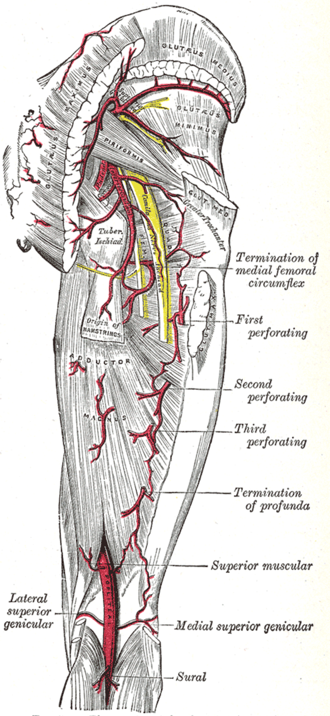
Adductor hiatus
The Adductor Hiatus
The adductor hiatus, also known as the hiatus magnus, represents a significant anatomical landmark within the human thigh. It is defined as a gap or opening that occurs between the posterior border of the adductor magnus muscle and the linea aspera of the femur. This specific discontinuity is not merely a passive space but an active conduit, serving as the terminal point of the adductor canal.
Its primary and most critical function is to allow the passage of the femoral artery and vein from their position in the anterior compartment of the thigh to the posterior compartment. This transition is fundamental for the continued vascular supply to the lower limb as these vessels transform into the popliteal artery and vein upon entering the popliteal fossa.
Vascular Dynamics and Clinical Implications
The adductor hiatus is a crucial nexus for the vascular system of the lower extremity. As the femoral artery and vein navigate through this opening, they are essentially rerouted from the anterior thigh to the popliteal region. This anatomical arrangement is vital for maintaining adequate perfusion to the muscles of the posterior thigh, the knee joint, and the entire lower leg.
Clinically, the adductor hiatus is of considerable importance. It is a landmark for surgical procedures, including vascular interventions and orthopedic surgeries around the knee and distal thigh. Surgeons must possess a thorough understanding of its precise location and the structures passing through it to avoid iatrogenic injury.
Furthermore, conditions such as deep vein thrombosis (DVT) can involve the vessels at or near the hiatus, and its anatomical configuration can influence the spread and detection of such thrombotic events. The potential for entrapment of the popliteal artery or vein within or adjacent to the hiatus, though rare, also underscores its clinical relevance.
Embryological Development and Anatomical Variation
The formation of the adductor hiatus is intrinsically linked to the complex embryological development of the lower limb. During fetal development, the vascular structures and musculature undergo significant remodeling. The adductor magnus muscle develops from the posterior compartment musculature but also contributes to the medial compartment, and its inferior portion forms the boundaries of the hiatus.
While generally consistent, there can be minor anatomical variations in the size, shape, and exact location of the adductor hiatus among individuals. These variations, though typically subtle, can sometimes influence surgical planning or the presentation of vascular pathologies. The consistent presence of this hiatus across the human population highlights its evolutionary significance, likely related to optimizing blood flow and structural integrity for bipedal locomotion.
Biomechanics and Functional Significance
From a biomechanical perspective, the adductor hiatus plays a role in the functional integrity of the leg. The adductor magnus muscle itself is a powerful adductor of the thigh, and its inferior fibers contribute to knee flexion and extension. The presence of the hiatus allows the major vascular supply to reach the posterior structures without being unduly compressed or kinked during the dynamic movements of the leg.
This ensures that muscles can receive oxygen and nutrients efficiently, even during strenuous activity. The relationship between the adductor hiatus and the adductor tubercle on the femur also provides a stable anatomical reference point. The distance from the hiatus to the tubercle, typically 8–13.5 cm superiorly, is a consistent measurement used in anatomical descriptions and clinical assessments, aiding in the precise localization of structures within the thigh.
Beyond the Hiatus
The adductor hiatus is part of a larger anatomical network. It is the terminal opening of the adductor canal, also known as the Hunter's canal, a fascial tunnel that begins in the mid-thigh and extends to the hiatus. This canal protects the femoral vessels and saphenous nerve as they travel distally.
Upon exiting the hiatus, the vessels enter the popliteal fossa, a diamond-shaped space behind the knee that houses the popliteal artery, popliteal vein, and the tibial and common fibular nerves. Understanding the adductor hiatus necessitates an appreciation of these contiguous anatomical regions and the continuous flow of structures through them. The interplay between muscle, bone, and vascular pathways at the adductor hiatus exemplifies the intricate and interconnected nature of human anatomy.
See also
Frequently Asked Questions
What is the adductor hiatus?+
Why does the adductor hiatus matter for blood flow?+
Where is the adductor hiatus located?+
How do doctors use the adductor hiatus?+
Can the adductor hiatus cause health problems?+
Based on content from Wikipedia · Licensed under CC BY-SA 4.0